MRI is a crucial tool for diagnosing back pain and spine issues. It offers detailed images of the spine that help doctors identify conditions like herniated discs, spinal stenosis, and fractures. Unlike X-rays or CT scans, MRI provides a more comprehensive view of soft tissues, spinal cord, and nerves, aiding in accurate diagnosis. Doctors can create tailored treatment plans through physical therapy, medication, or surgery. Consulting a skilled back doctor with MRI scans can significantly improve diagnosis and treatment outcomes for spine-related problems.
Understanding The Importance Of Accurate Diagnosis
Accurate diagnosis is crucial for managing back pain effectively, as the spine consists of various components such as bones, discs, nerves, and soft tissues. Identifying the underlying cause is essential to avoid misdiagnosis and inappropriate treatments that could worsen the condition. Without a precise diagnosis, patients may experience frustration and prolonged pain. An accurate diagnosis enables back doctors to choose the most effective treatment, set realistic recovery expectations, and prevent future complications. Early detection through advanced imaging like MRI can help avoid chronic pain or permanent damage, ensuring patients receive the proper care.
What Is An MRI and How Does It Help Diagnose Spine Issues?
Magnetic Resonance Imaging (MRI) is a noninvasive diagnostic tool that uses powerful magnets and radio waves to generate detailed images of internal structures. It focuses on soft tissues like muscles, ligaments, nerves, and intervertebral discs. MRI complements X-rays and CT scans in diagnosing spine-related conditions.
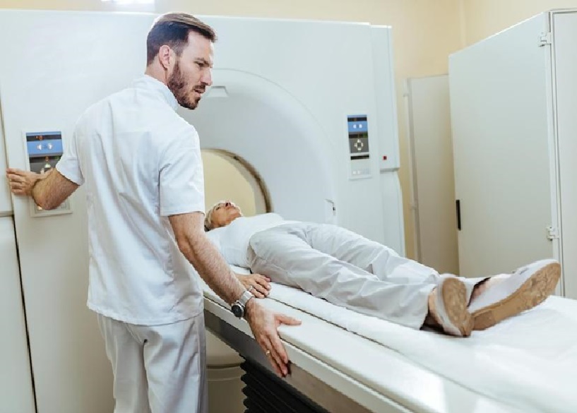
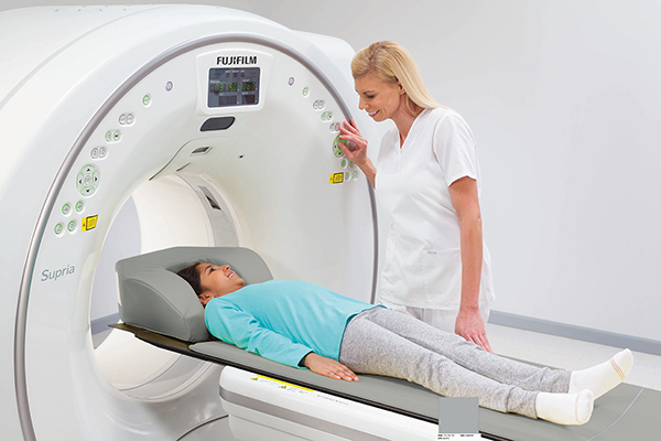
During the procedure, patients lie on a table that moves into the MRI machine, with 30 to 60 minutes of imaging sessions. At Tellica Imaging, MRIs are vital for identifying herniated discs, spinal stenosis, tumors, infections, and inflammation. This technology provides critical insights for accurate diagnoses and personalized treatment plans.
Benefits Of Using MRI for Diagnosing Spine Issues

MRI offers several advantages for spine diagnostics, notably providing high-resolution images without exposing patients to ionizing radiation. This is especially beneficial for patients who require frequent imaging, as MRI can be performed multiple times without the long-term risks associated with repeated X-rays or CT scans.
MRI also provides a comprehensive spine view, including soft tissues such as muscles, ligaments, intervertebral discs, and the spinal cord. This allows doctors to accurately assess conditions like herniated discs and their impact on nerve roots, which can influence treatment decisions.
Additionally, MRI can establish a baseline for spine health and monitor chronic conditions over time. Regular scans help track disease progression, evaluate treatment effectiveness, and adjust care plans, ultimately improving patient outcomes and quality of life.
Common Spine Issues Diagnosed Through MRI
MRI is essential in diagnosing common spine issues, such as herniated discs, spinal stenosis, and spinal tumors. A herniated disc can press on nerves, and MRI helps locate the herniation, guiding treatment like physical therapy or surgery. Spinal stenosis, causing nerve compression and leg pain, is also assessed by MRI to determine treatment, including possible surgery. MRI is crucial for detecting spinal tumors, infections, and inflammatory conditions, enabling early intervention and effective treatment to prevent complications and aid recovery.
How Back Doctors Interpret MRI Results
Interpreting MRI results requires specialized expertise from back doctors, such as radiologists or orthopedic specialists. They assess the images for issues like disc herniations, spinal misalignments, and degeneration, considering the patient’s symptoms and history for a comprehensive diagnosis. The process involves evaluating image quality, vertebral alignment, disc condition, and nerve compression or inflammation signs. A detailed report guides treatment planning and may lead to additional imaging or follow-up MRIs, especially for chronic conditions. Close communication ensures timely interventions and optimal patient outcomes.
Advancements In MRI Technology For Spine Diagnosis
Advancements in MRI technology have significantly enhanced spine diagnostics. High-field MRI systems provide higher-resolution images and faster scanning, improving the detection of subtle abnormalities. Innovations like functional MRI (fMRI) and diffusion tensor imaging (DTI) offer insights into pain perception and spinal cord integrity, aiding in understanding complex pain syndromes. Additionally, advanced image analysis software allows for better tracking of spinal changes over time, improving pre-surgical planning and surgical outcomes. These ongoing developments equip back doctors with the tools for more accurate diagnoses and tailored treatments.
Other Diagnostic Tools Used In Conjunction With MRI
MRI is often used alongside other diagnostic tools to view spine issues comprehensively. X-rays are commonly the first step to assess spinal alignment and rule out fractures, focusing on bony structures. For more detailed bone imaging, CT scans identify fractures or spinal deformities, and CT myelograms may enhance spinal cord and nerve root visualization. Additionally, electromyography (EMG) and nerve conduction studies assess nerve function, complementing MRI results. By combining these diagnostic methods, doctors can more accurately diagnose spine conditions and determine appropriate treatment.
The Role Of MRI In Treatment Planning For Spine Issues
After interpreting MRI results, back doctors develop personalized treatment plans based on the severity and extent of the condition. This may involve conservative options like physical therapy and pain management or, if necessary, surgical interventions. For example, a herniated disc causing nerve compression may be treated with medicine, medications, and possibly injections. If conservative methods fail, surgery such as discectomy or spinal fusion may be considered. MRI results also help set realistic recovery expectations and guide ongoing monitoring, ensuring effective treatment adjustments for optimal outcomes.
Conclusion: The Significance Of MRI in Diagnosing Spine Issues And Improving Patient Outcomes
In conclusion, MRI has transformed spine diagnosis and management by providing detailed views of soft tissues, discs, and nerves. This enables back doctors to create effective treatment plans, improving patient outcomes and quality of life. As MRI technology advances, doctors are better equipped to diagnose and treat various spine conditions. MRI offers a comprehensive approach to spine health combined with other diagnostic tools. Patients experiencing back pain should consult qualified back doctors who prioritize MRI for early detection and accurate diagnosis, ensuring timely treatments and promoting recovery for a healthier, more active lifestyle.


